|

Sometimes we’re presented with a dog that is kicking his leg out in an odd way when running. Sometimes we’ll see a dog that walks normally for the most part, but will occasionally hold up a back leg for a step or two, extend it backward, and then resume normal walking. Both of these scenarios are suggestions for us to examine the hind legs, and in particular the kneecaps, or patellas. Still other times, we’re presented with a dog that appears normal at home, and we find something unusual with their stifles on routine examination.
Dislocating kneecaps, or medial luxating patellas, are one of the more common orthopedic abnormalities that we find in our canine patients. The smaller breeds are over represented, though we find this in some of the larger breeds like Labrador Retrievers.
You can feel your own patellas by gripping the front of your knee with your thumb and index finger. If your leg is straight and relaxed, you can wiggle your patella back and forth ever so slightly.
WARNING: ANATOMY LESSON FOLLOWS WITHIN THE NEXT PARAGRAPH!!!
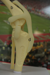
Your dog’s knee is called the stifle, and it is set up very similarly to yours. Your patella sits in a groove (a) at the end of your thigh bone (femur, b). This groove ought to be in the middle of a straight line connecting the point of the hip (cranial ventral iliac spine, c) to the boney knob on the front of the top of the shin bone (tibial tuberosity, d). This alignment of the boney structures is important, because the patella is connected to the thigh muscles above (which originate at the point of the hip, c) and the patellar ligament below (which connects to the tibial tuberosity, d).
Whew! You made it through the tough part, which leads me to…
THE TAKE-HOME POINT: All of the anatomic structures related to the kneecap ought to line up very nicely in a straight line.
THE PROBLEM: This doesn’t always happen.
Sometimes the groove is too shallow. Sometimes the femur is bowed. Most of the time the tibial tuberosity isn’t centrally located on the shin bone but rather it is located more to the inside aspect of the shin bone. A dog has his/her mom & dad to thank for any and all of this. This isn’t an injury or a result of being too active, it’s a result of how the bones grew way back when.
Luxating patellas, dislocating kneecaps, are graded on a scale of 1 through 4.
Most dogs have a grade of 0. The kneecaps are well in place, and nothing short of a sledge hammer is going to make them pop out of place. For the abnormals…
- Grade 1: I can push the kneecap out with my thumb, but as soon as I release my thumb’s pressure, the kneecap will return to its correct position. Grade 1 dogs are rarely lame.
- Grade 2: I can push the kneecap out with my thumb, but when I release my thumb’s pressure, the kneecap does not return quickly. It will return to its correct position as the dog moves around. Arthritis formation will occur with these dogs, and surgery to correct the luxation prior to significant arthritis formation is advised as a preventive step.
- Grade 3: The kneecap can dislocate on its own with no outside force or encouragement. I am able to replace it with my thumb and index finger, and it will remain in place… until it pops out again. For many of these dogs, the kneecap is out of place more often than it’s in place, and this is not good from an arthritis standpoint. The kneecap rubs the end of the thigh bone in a way that it was not meant to do. Dogs may not show overt signs of outward pain, but this abnormal friction is painful and will lead to additional painful consequences. These dogs need surgery.
- Grade 4: The kneecap isn’t living where it is supposed to, and I am unable to replace it manually. These dogs need surgery.
There are several surgical techniques that are employed when correcting a medial patellar luxation. Dr. Doug has been performing these techniques since the late ’70’s, and I have learned from him. After seeing patient after patient recover wonderfully from surgery and experience the comfort that an appropriately functioning knee provides, I’ve come to often say to clients, “If I had to choose an orthopedic surgery that my dog *had* to experience, it would be correction for medial patellar luxation.”
Of course our patients are blanketed with appropriate pain medication before, during and after surgery, and it is important for our pet owners to follow our post-operative instructions very closely. Most of our patients are walking very well by the time of suture removal at 2 weeks post-op. Once in a blue moon a dog must be retrained to used the leg, and we have some creative ways of achieving this. Close to 100% of dogs return to normal function within a few weeks of surgery, and then life goes very well.
We’ve been happy with how our patients have responded to surgical correction, and if your dog has a luxating patella and you’re considering surgery, please don’t hesitate to raise any of your questions with us.