 Stepping into our blog spotlight today is… our microscope. Nearly all of us have used a microscope at some point in our lives. For most it was during a high school biology class. You may have seen an amoeba or plant cells or something in between. If you were fortunate enough to have a toy microscope as a kid, you certainly enjoyed making all sorts of things get bigger before your very eyes, from ants to food to your fingerprint.
At the Cuyahoga Falls Veterinary Clinic, we use our microscope every day for a variety of health care areas. I’ll itemize the common areas, and you can click on the photos for more details.
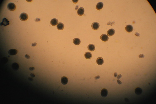
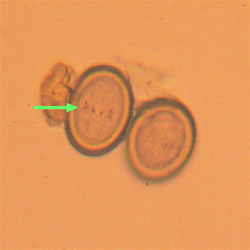
The most common use of our microscope is for evaluating fecal samples of dogs and cats as we screen them for intestinal parasites. We’e talked about the importance of keeping our pets parasite-free both for their protection and for ours. Looking for microscopic parasite eggs helps us identify the species of parasites that are present.
  |
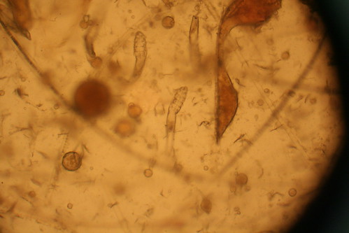
We also use the microscope to look for organisms such as yeast or bacteria. It’s not uncommon for dogs and cats to develop ear infections, and to choose the best therapy as well as set a baseline for future evaluation, we’ll take a sample of the “gunk” in the ear and look at it under the microscope. Yeast cells have a characteristic appearance while bacteria can have a couple of different appearances, depending on the type that’s present.
 |
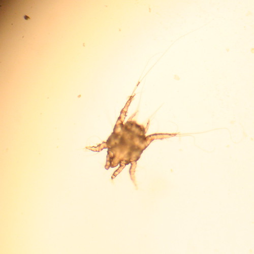
Once in a while we’ll see external parasites under the microscope, like demodectic mange or ear mites. Finding these helps us to choose the most effective treatment plan for our patient. Demodex can be very hard to find, even when we’re quite sure it’s present on the patient, but when it’s found, the diagnosis is a no-doubter.
  |
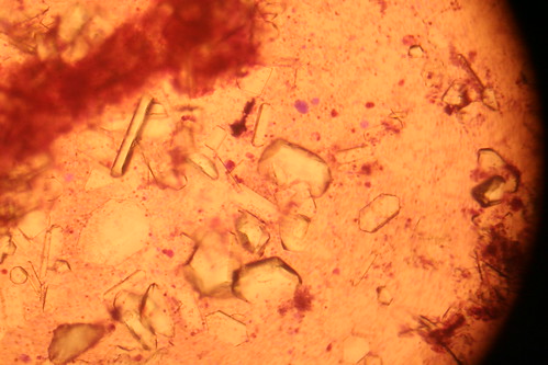
If a cat or dog is having trouble urinating or is showing blood in the urine, we’ll evaluate the urine under the microscope, looking for unusual cells or urinary crystals to explain the symptoms.
 |
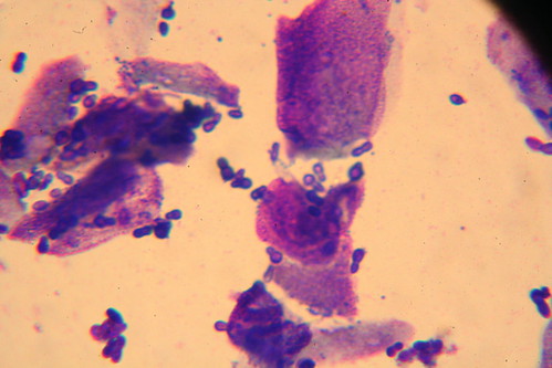
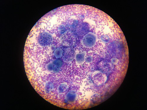
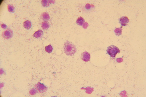
We’ll use the microscope to look at cell samples from unusual growths. This particular dog had swellings on the face where lymph nodes usually hide. Aspirating cells from these and sending them to a pathologist for him to use his microscope gave us a diagnosis. In this first case, squamous cell carcinoma was at play, which is not good form of cancer. Below that is an example of a Mast Cell Tumor aspirate, an indication for surgery.
|
With all of the advances in medical technology, it’s important for good medical professionals to avoid leaving behind tried and true diagnostic tools such as a good microscope. We couldn’t get through a day without ours.